


Advancements in Regenerative Medicine: The Bioengineering of Functional Esophageal Tissue
The landscape of regenerative medicine has reached a pivotal milestone as a consortium of United Kingdom-based scientists announced the successful development and transplantation of fully functioning bioengineered food pipes, or esophaguses, into porcine models. This breakthrough, spearheaded by researchers at Great Ormond Street Hospital (GOSH), University College London (UCL), and the Francis Crick Institute, represents a significant leap toward the realization of lab-grown organs for human clinical use. By utilizing a sophisticated combination of tissue engineering and stem cell biology, the team has addressed one of the most complex challenges in gastroenterology: the replacement of esophageal tissue that has been damaged by congenital defects, trauma, or oncological interventions.
For decades, patients suffering from severe esophageal conditions,such as esophageal atresia in infants or esophageal cancer in adults,have been forced to rely on invasive “stomach pull-up” procedures or intestinal interpositions. These traditional surgical methods are fraught with complications, including high rates of infection, strictures, and long-term nutritional deficiencies. The shift toward bioengineered solutions promises a paradigm where the body’s own cellular material is used to reconstruct vital organs, thereby eliminating the risk of immunological rejection and the lifelong necessity for immunosuppressant drugs. This recent success in mini-pigs, whose physiology closely mirrors that of humans, provides the critical preclinical validation required to move into human trials.
The Bioengineering Paradigm: Decellularization and Recellularization
The technical foundation of this achievement rests on a two-stage process involving decellularization and recellularization. In the initial phase, researchers utilize donor esophageal tissue,often from animal sources or deceased human donors,and apply a rigorous chemical and physical process to strip away all living cells. What remains is an extracellular matrix (ECM), a protein-based “scaffold” that retains the intricate structural geometry and mechanical properties of the original organ. This scaffold is crucial because it provides the necessary biochemical cues for new cells to adhere and differentiate, while being entirely non-immunogenic because the cellular components that trigger a host’s immune response have been removed.

The second, more complex phase involves seeding this scaffold with a patient’s own stem cells. In this specific study, the UK team utilized mesenchymal stem cells and epithelial cells. These cells were cultured within a specialized bioreactor,a controlled environment that mimics the physiological conditions of the human body, providing nutrients, oxygen, and mechanical stimulation. Over several weeks, these cells populated the scaffold, organizing themselves into the distinct layers required for esophageal function, including the inner mucosal lining and the outer muscular layers. The ability of these cells to not only survive but to organize into complex tissue structures marks a major victory for the field of synthetic biology.
Preclinical Validation and Physiological Integration
The transition from a laboratory setting to a living organism is the most significant hurdle in medical innovation. The successful transplantation of these bioengineered pipes into mini-pigs served as a rigorous proof-of-concept. Post-operative monitoring revealed that the engineered grafts integrated seamlessly with the animals’ existing tissues. Crucially, the researchers observed the development of a functioning vascular network,a process known as angiogenesis. Without the growth of new blood vessels to supply oxygen and nutrients to the graft, the engineered tissue would undergo necrosis and fail.
Beyond simple integration, the grafts demonstrated functional efficacy. The study reported that the engineered esophaguses were capable of peristalsis, the rhythmic muscular contractions required to move food and liquid through the digestive tract. This functional success is attributed to the presence of both smooth muscle cells and neural progenitor cells within the graft, which allowed for the eventual integration of the host’s nervous system. The longitudinal data collected from these porcine models indicate that the grafts are durable and grow in tandem with the animal, a factor that is particularly vital for pediatric applications where the organ must expand as the child matures.
Market Implications and the Path to Clinical Translation
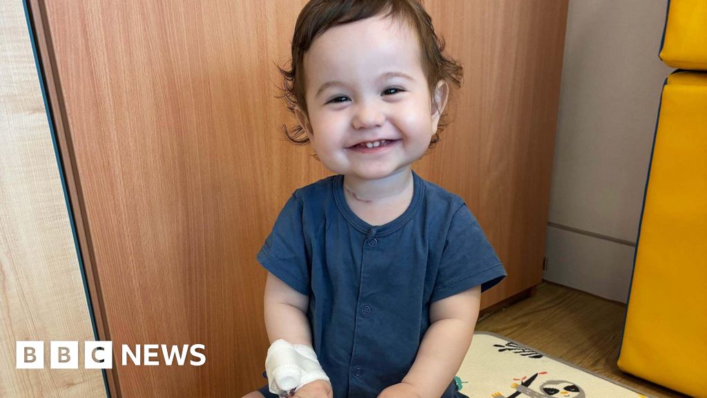
The successful pilot in porcine models has cleared the regulatory path for the first-in-human clinical trials, which are expected to target pediatric patients with limited surgical options. From a business and healthcare infrastructure perspective, the implications are profound. The current cost of treating chronic esophageal conditions is astronomical, factoring in multiple corrective surgeries, long-term hospitalizations, and the management of complications. A one-time, lab-grown curative intervention could significantly reduce the long-term financial burden on national healthcare systems and private insurers alike.
Furthermore, this development signals a burgeoning sector in the biotechnology market: the “organ-on-demand” industry. While the esophagus is a relatively simple tube compared to the heart or liver, its successful engineering provides a blueprint for the reconstruction of other hollow organs, such as the trachea, bowel, and bladder. As the technology scales, the focus will shift toward the automation of the bioreactor process to ensure consistency, safety, and cost-effectiveness. The involvement of top-tier UK research institutions underscores the country’s position as a global leader in life sciences, attracting significant venture capital and government interest into the field of regenerative medicine.
Concluding Analysis: A New Era of Personalized Surgery
The achievement of UK scientists in growing and transplanting functional food pipes is more than a localized medical success; it is a harbinger of a broader transformation in the medical sciences. We are moving away from the “one-size-fits-all” approach of mechanical prosthetics and toward a personalized, biological approach to surgery. The use of a patient’s own cells to grow an organ eliminates the ethical and logistical hurdles associated with traditional organ donation, such as donor shortages and the bioethical complexities of xenotransplantation.
However, as we move toward human trials, rigorous oversight remains paramount. The long-term stability of these grafts in humans, potential oncogenic risks of rapid stem cell proliferation, and the scalability of the manufacturing process are questions that must be addressed. Nevertheless, the successful integration of bioengineered tissue in a large animal model provides the strongest evidence to date that lab-grown organs are no longer a theoretical ambition but a clinical reality. As this technology matures, it will likely redefine the standard of care for millions of patients worldwide, offering a permanent solution where previously there were only temporary fixes.


